
Fig. 4.
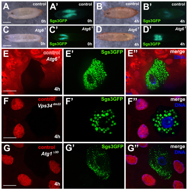
Loss of Atg6 leads to defects in protein secretion. (A,A′) Control pupa (0 hour) showing expression of SgsΔ3-GFP in salivary glands (n=10). (B,B′) Four hours later the same pupa as in A lacked SgsΔ3-GFP. (C-D′) Homozygous Atg61 mutants possess SgsΔ3-GFP protein at 0 hour (C-C′) (n=12), but are unable to secrete it 4 hours after puparium formation (D-D′). (E-E″) Control cells (mCherry positive) are able to secrete Sgs3Δ3-GFP whereas homozygous Atg61 mutant cells (mCherry negative) retain SgsΔ3-GFP in the cytoplasm (n=12). (F-F″) Control cells (mCherry positive) secrete SgsΔ3-GFP whereas Vps34dm22 mutant cells retain SgsΔ3-GFP granules in the cytoplasm (n=11). (G-G″) Control cells (mCherry positive) secrete SgsΔ3-GFP whereas Atg1Δ3D expressing cells retain SgsΔ3-GFP granules in the cytoplasm (n=10). Scale bars: 1 mm in A,C; 50 μm in E-G.