
Fig. 5.
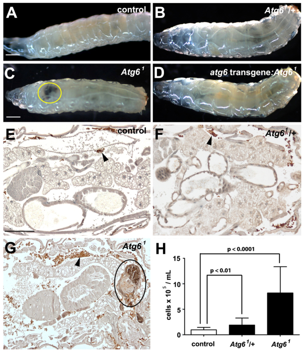
Loss of Atg6 leads to melanotic blood cell mass formation. (A-D) White-light images of wandering third instar larvae. Parental control w1118 (A) and heterozygous Atg61 (B) larvae are phenotypically similar. Homozygous Atg61 mutant larvae (C) contain melanotic blood cell masses (yellow ring), and this phenotype is rescued by ubiquitous expression of a GFP-Atg6 transgene (D). (E-G) Hemocytes were visualized by immunohistochemistry in third instar larvae expressing GFP, driven by hmlΔ-GAL4 (arrowheads). Histological sections of w1118 (E) and Atg61 heterozygous (F) larvae contain few hemocytes (brown) and no visible melanotic masses, whereas sections of homozygous Atg6 mutant larvae (G) reveal many hemocytes, which surround melanotic masses (black ring). (H) Quantification of hemocytes from w1118, Atg61/+ and Atg61/Atg61 larvae revealed a significant increase in hemocyte number in both heterozygous and homozygous Atg6 mutant larvae compared with control w1118 larvae. For each genotype, n=20 animals. Error bars represent s.e.m. A one-tailed t-test was used for statistical analysis and P-values relative to w1118 are: Atg61/+, P=0.004; Atg61/Atg61, P=2.6×10⁻⁶. Scale bars: 500 μm in A,C; 100 μm in E.