
Fig. 7.
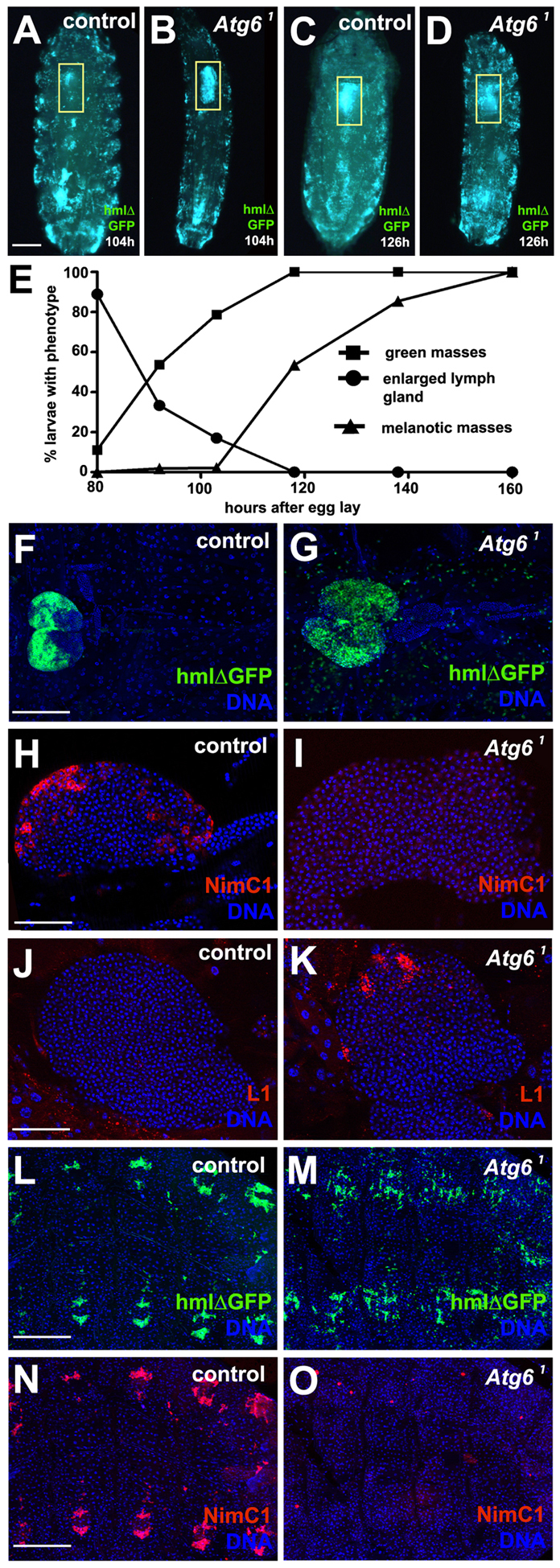
Loss of Atg6 leads to enlargement of lymph gland and altered blood cell development. (A) Control larva at 104 hours after egg lay expressing GFP in blood cells. (B) Homozygous Atg61 mutant larva at 104 hours after egg lay expressing GFP in blood cells. Note the enlarged lymph gland (yellow box). (C) Same control animal as in A at 126 hours after egg lay has formed a prepupa. (D) Same Atg61 mutant animal as in B at 126 hours after egg lay did not pupariate, and appeared to have an increased number of circulating blood cells. (E) Graph showing progression of different phenotypes exhibited by Atg6 mutants during larval development. The larval stage-specific numbers of teeth on mouth hooks of control and Atg6 mutant larvae were used to normalize development of these genotypes. As larvae progress through development there is an increase in formation of blood cell aggregates followed by melanotic masses (n=157). (F) Control hmlΔ-GAL4 UAS-GFP lymph gland at third instar larval stage (n=10). (G) Atg61 mutant hmlΔ-GAL4 UAS-GFP lymph gland at third instar stage (n=10). (H) Control third instar lymph gland stained for NimrodC1 (NimC1) showing expression in the cortical zone (n=7). (I) Atg61 mutant third instar lymph gland stained for NimC1 showing a complete lack of expression in the cortical zone (n=15). (J) Control third instar larval lymph gland stained for lamellocyte specific antigen L1 showing no expression in the cortical zone (n=7). (K) Atg61 mutant third instar animal lymph gland with increased expression of L1 in the cortical zone (n=7). (L) Control hmlΔ-GAL4 UAS-GFP animal showing sessile blood cells that are located in a reiterated pattern along abdominal segments (n=7). (M) hmlΔ-GAL4 UAS-GFP-expressing Atg61 mutant animals possess less patterned sessile blood cells along the abdominal segments than controls (n=7). (N) NimC1 staining of animal shown in L. (O) NimC1 staining of animal shown in M indicates that this blood cell antigen is missing in sessile blood cells. Yellow boxes in A-D delineate lymph glands. Scale bars: 250 μm in A; 100 μm in F; 50 μm in H,J; 200 μm in L,N.