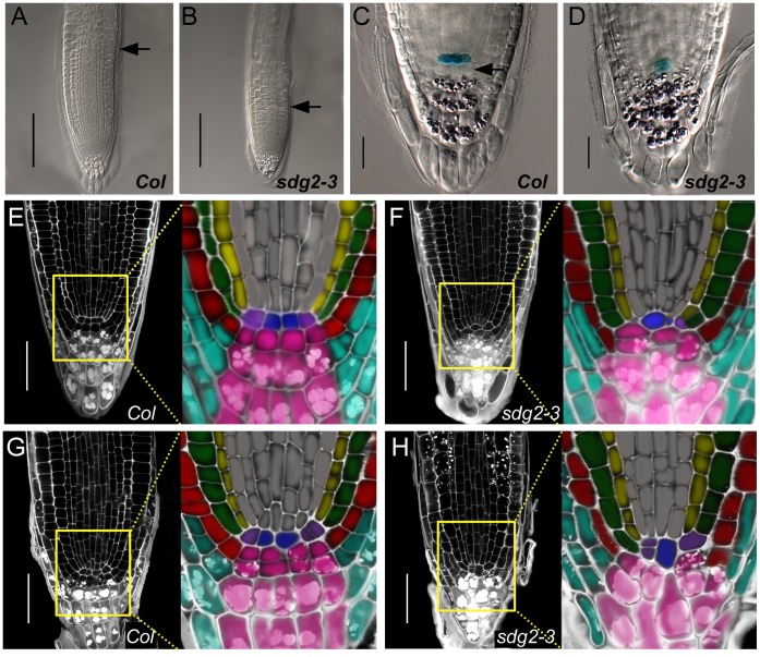
Figure 3. Loss of SDG2 impairs the primary root stem cell niche maintenance. A.
and B, Comparison of primary root apical meristem sizes between wild-type Col and the mutant sdg2-3, respectively. DIC images were taken on the roots of 6-day-old seedlings. Arrowheads indicate positions of the transition from meristem to elongation zone. Bar = 100µm. C and D, Comparison of QC25:GUS expression and root cap cell layer organization between Col and sdg2-3, respectively. DIC images were taken on GUS- and Lugol-stained root tips of 6-day-old seedlings. Arrowheads indicate the columella initial cell layer. Bar = 20 µm. E and F, Comparison of cell layer organization of root apical meristem between Col and sdg2-3, respectively. Confocal images were taken on PI-stained roots of 6-day-old seedlings. Bar = 50 µm. The close-up regions are shown by color indication of different cell types: QC cell in blue, columella root cap and columella initial cells in rose, lateral root cap cells in sky-blue, epidermal cells and epidermis/lateral root cap initials in red, cortex cells in green, endodermal cells in yellow, cortex/endodermis initials in purple, stele cells and stele initials in gray. G and H, Comparison of cell layer organizations of root apical meristem between Col and sdg2-3, respectively. Confocal images were taken on PI-stained roots of 14-day-old seedlings. Bar = 50 µm. The close-up regions are shown with colorations as described in E and F.

