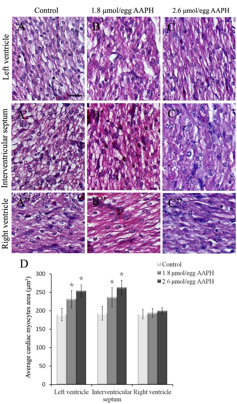
Figure 8. Effect of AAPH on cardiac myocytes size of chick embryo.
(A–C) H&E-staining of heart sections obtained from left ventricle of HH 40 stage chick embryo of (A) control, (B) 1.8 µmol/egg AAPH and (C) 2.6 µmol/egg AAPH group. (A’–C’) H&E-staining of interventricular septum from HH 40 stage chick embryo of (A’) control, (B’) 1.8 µmol/egg AAPH and (C’) 2.6 µmol/egg AAPH group. (A’’–C’’) H&E-staining of right ventricle from HH 40 stage chick embryo of (A’’) control, (B’’) 1.8 µmol/egg AAPH and (C’’) 2.6 µmol/egg AAPH group. (D) Statistical data of average cardiac myocytes surface area. Results presented as mean ± S.D (n = 10) calculated by SPSS13.5 software, *p<0.05. Scale bar = 20 µm in A–C, A’–C’, A’’–C’’.

