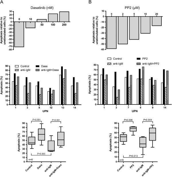
Figure 6.
PP2 and dasatinib suppress BCR-induced cell survival. (A) Primary MCL cells (UPN3) were either left untreated or stimulated for 24 h with an anti-IgM antibody in the presence or in the absence of various concentrations of dasatinib (10 to 200nM) (top panel). Apoptosis rates (Annexin V +/PI+ cells) were measured by flow cytometry after gating on CD19+ cells. For each BCR-stimulated condition (± dasatinib), the percentage of apoptotic cells was normalized to unstimulated cells and calculated as follows: [(% apoptosis BCR stimulated cells -% apoptosis BCR-unstimulated cells) / (% apoptosis BCR-unstimulated cells)] x100. Middle panel: Apoptosis rates from 6 MCL cases were measured from unstimulated or BCR-stimulated cells (24 h) either in absence or presence of 10 nM dasatinib. All measurements were done in duplicate and the mean is provided. Results are also shown as median ± quartile (box) ± SE (bars) (n=6) (bottom panel). Differences between groups were determined using the paired Student t test. (B) Primary cells were treated with PP2 according to the same protocol described in (A).

