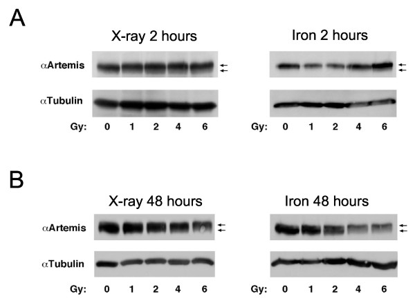
Figure 3.
Artemis phosphorylation is delayed following high LET damage. Western blots were probed for total Artemis protein and mobility shifts of the protein indicate phosphorylation has occurred. Blots are shown 2 h post X-ray or iron nuclei exposure (A, left and right panel respectively) and 48 h post X-ray or iron nuclei exposure (B, left and right panel respectively). Arrows indicate location of basally-phosphorylated Artemis (lower arrow) and hyper-phosphorylated Artemis (upper arrow).

