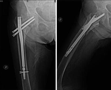
Fig. 3.

Six-month follow-up radiographs show nonunion of the intertrochanteric fracture and breakage of the nail. Note the sliding of the cephalic screws and the failed sliding of the nail around the distal screw. On the lateral view, the tip of the nail is abutting the anterior cortex
