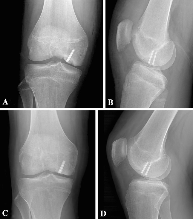
Fig. 7A–D.

Radiographs at (A) age 15 years 9 months (immediately postoperatively) and (B) 1 week postoperatively confirm fragment fixation of the MFC OCD with the two variable-pitch screws. The trochlear OCD remains visible. Followup radiographs at (C) age 16 years 5 months and (D) 16 years 8 months demonstrate healing of the MFC OCD lesion, with the boundary between the progeny fragment and parent bone nearly nondistinct. In addition, the radiodensity of the subchondral bone of the trochlear OCD is the same as the surrounding parent bone.
