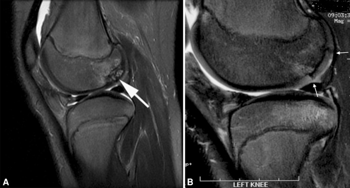
Fig. 2A–B.
(A) Corresponding T2 fat-saturated MR image in sagittal view shows a large OCD defect at the posterior portion of lateral condyle (arrow). (B) Corresponding T2 fat-saturated MR image in sagittal view 28 months postallografting shows allograft incorporating well with the host site without bony gap and persistent demarcation between allograft and native cartilage (arrow).

