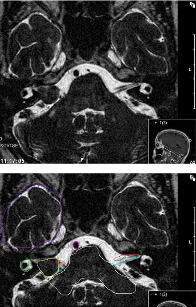
Figure 1.

Axial T2-weighted MRI with a still visible CSF-interface between tumour and brain. The largest diameter of the tumour in the CPA cistem is 14 mm. Yellow: vestibular schwannoma; green: labyrinth; red: ipsilateral and contralateral facial nerve; blue: ipsilateral and contralateral vestibulocochlear nerve; white: brainstem and cerebellar peduncle; purple: caudal temporal lobe; pink: basilar artery.
