
Fig 8.
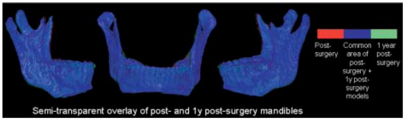
Semitransparent overlay of registered 1-week and 1-year postsurgery mandibular models of patient in Fig 7. Other anatomic structures are masked for better visualization of changes in mandible. Red, presurgery model; blue, area where pre- and postsurgery models overlap; green, postsurgery model.