Fig 8.
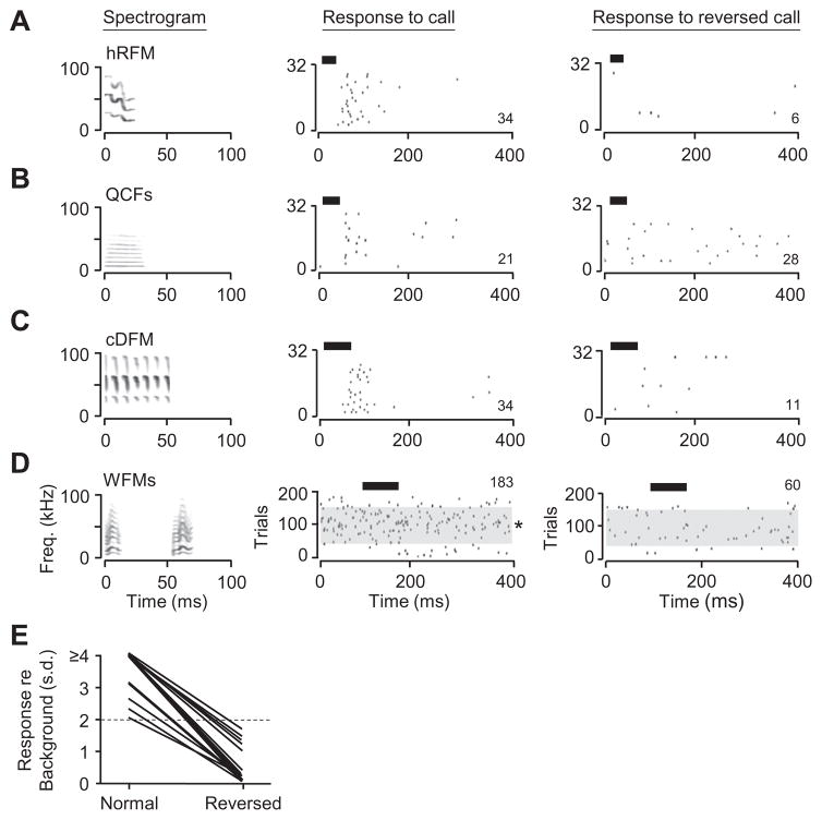
Time-reversed social calls did not elicit strong responses in amygdalar neurons. (A–C) Responses of one neuron to three normal and time-reversed calls. For each call, the locked discharge was reduced. (D) Persistent spike discharge of a different neuron was substantially reduced when WFMs call pair was reversed. The raster shows spikes during 200 trials; see Fig. 2 for description. For the normal call, asterisk indicates that the response to stimulus presentation was statistically greater than during the no-sound trials (*p < 0.05, t-test). For the reversed call, there was no statistical difference in firing rate for sound vs. no-sound trials. All sounds in (A–D) were presented at 20-dB attenuation. (E) Response of neurons to normal and time-reversed calls. Response is computed as the number of standard deviations (s.d.) above baseline spiking activity. Values above two standard deviations (dashed line) represent our criterion for a response. In all cases, spike discharge was substantially reduced with time-reversal of calls.

