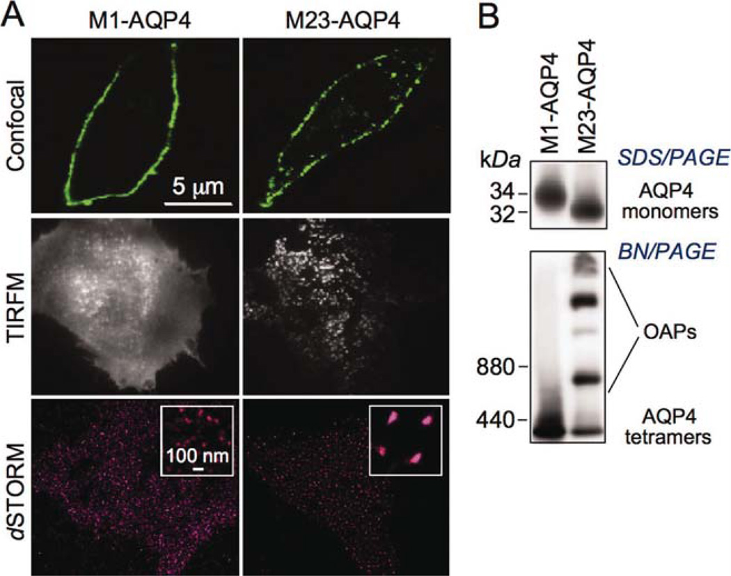
Fig. 1.
Characterization of AQP4-transfected CHO cells. (A) Immunofluorescence of CHO cells stably transfected with M1- or M23-AQP4, stained with anti-C-terminus AQP4 antibody and fluorescent secondary antibody, and imaged by confocal microscopy (top), TIRFM (middle), and dSTORM (bottom). (B) AQP4 immunoblot of cell homogenate following SDS/PAGE (top) and (nondenaturing) BN/PAGE (bottom). [Color figure can be viewed in the online issue, which is available at wileyonlinelibrary.com.]

