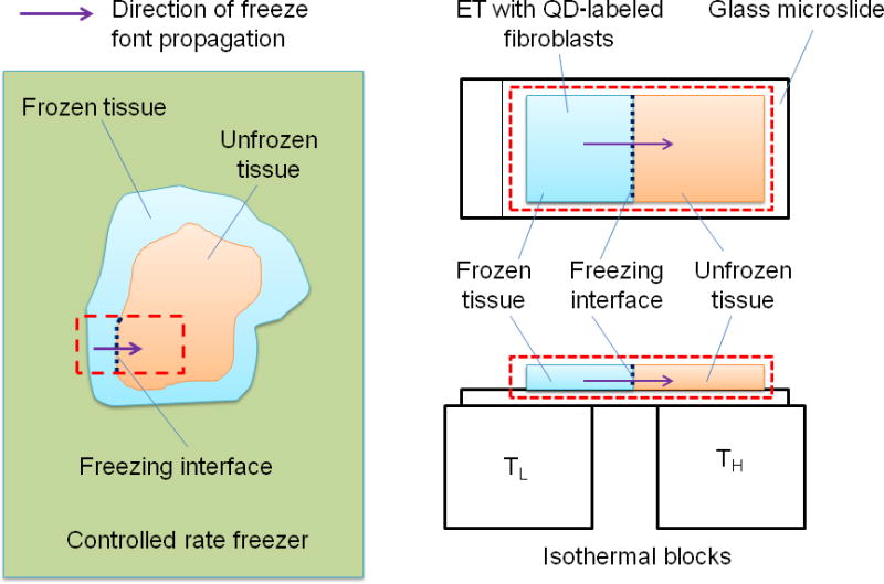
Fig. 1.

Schematic of the freezing procedure: The left side shows a typical procedure of tissue cryopreservation. The native/ engineered tissues are frozen inside a controlled-rate freezer. The freeze front propagates from the outside boundary to the interior of the tissue as shown by the bold arrow. This procedure is mimicked using a directional solidification stage. The tissue is placed on a glass microslide which rests on the cold and hot isothermal posts. Because of the temperature gradient created, the freeze front propagates along the direction shown by the bold arrow.
