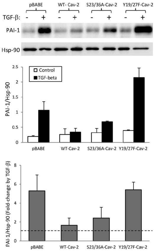
Fig. 4.

Effect of TGF-β on PAI-1 protein expression in Cav-2 KO MLECs re-expressing WT and phosphorylation-deficient mutants of Cav-2. WT-Cav-2, S23/36A-Cav-2, and Y19/27F-Cav-2 MLECs were plated at 3 × 10⁵ cells onto 150 mm dishes, 24 h later incubated without or with TGF-β (1 ng/ml) for additional 48 h, lysed and processed for SDS–PAGE and immunoblotting with indicated antibodies as described in Section 2. Top graph: Densitometric ratios of PAI-1/Hsp-90 from control (blank bars) and treated with TGF-β (solid bars) samples quantified based on the above immunoblots and expressed as Means ± S.D. (n = 3). Bottom graph: Fold-change by TGF-β relative to respective control samples expressed as Means ± S.D. (n = 3).
