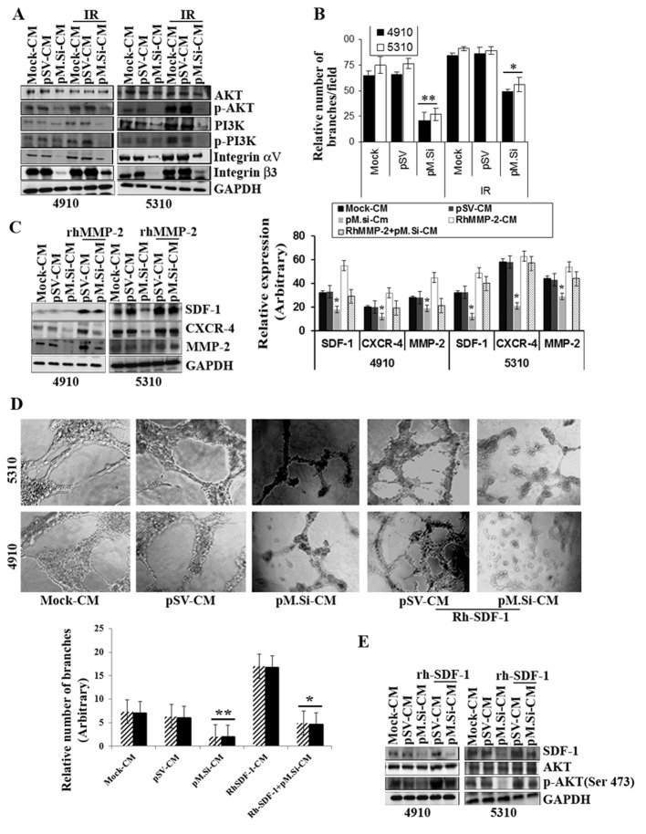
Figure 2.
pM.Si-CM inhibits IR-induced PI3K/AKT expression and angiogenesis in ECs and supplementation of rhMMP-/rhSDF-1 reverses inhibition. (A) ECs were grown in mock-, pSV- and pM.Si-CM with or without IR. The whole cell lysates were subjected to western blotting to check the expression levels of AKT, p-AKT, PI3K, p-PI3K, integrin αv and integrin β3 using specific antibodies. The blot was restriped and GAPDH was used as a loading control. (B) In vitro angiogenesis was done with overnight incubation of ECs at 37°C with mock-, pSV-, IR (8 Gy)-CM or pM.Si alone or in combination with IR (8 Gy). The cells were observed under a bright field microscope for the formation of capillary-like structures. The degree of angiogenic induction was quantified for the relative capillary length and number of branch points per field. The data are presented as the mean ± SE of three independent replicates with significance denoted by *p<0.05 and **p<0.01. (C) ECs from 4910 and 5310 cells were grown in the presence of mock-, pSV- or pM.Si-CM with or without IR (8 Gy) for 16 h. pSV- and pM.Si-CM were treated with 25 ng/ml rhMMP-2. Whole cell lysates of ECs were prepared at the end of 16-h treatment and subjected to western blotting to check the expression levels of SDF-1, CXCR4 and MMP-2 using specific antibodies. The blot was restriped and GAPDH was used as a loading control. The data are presented as the mean ± SE of three independent replicates with significance denoted by *p<0.05 and **p<0.01. (D) In vitro angiogenesis was carried out under similar conditions as noted in Fig. 1A and is representative of at least three independent repetitions. RhSDF-1 (25 ng/ml) was added to pSV- and pM.Si-CM and incubated for 16 h. The degree of capillary network formation is indicated in a graph. The data are presented as the mean ± SE of three independent replicates with significance denoted by *p<0.05 and **p<0.01. (E) Endothelial cells were treated with pSV-CM or pM.Si-CM supplemented with rhSDF-1 (25 ng/ml) for 16 h. The whole cell lysates were subjected to western blot analysis for the expression of SDF-1, AKT and p-AKT with their respective antibodies and GAPDH was used as the loading control.

