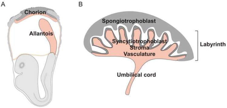
Figure 1.

Placenta structure at different times in development (a) Early stages of chorio-allantoic placenta formation. The placenta is formed from the fusion of the allantois with the chorion. The allantois is a mesodermal outgrowth emanating from the posterior primitive streak. It elongates and upon contact with the chorionic mesoderm gives rise to the labyrinth of the placenta. (b) Mouse placenta structure. The placenta consists of the several cell types and layers. The side of the placenta facing the fetus is the labyrinth. It consists of a vascular network contiguous with the umbilical cord. The network of vessels in the labyrinth form branched villous structures that are surrounded by mesenchymal stromal and syncytiotrophoblast cells. The maternal vessels run through the spongiotrophoblast region (facing the mother) to open into the intervillous space (white area) where physiologic exchange between mother and fetus occurs.
