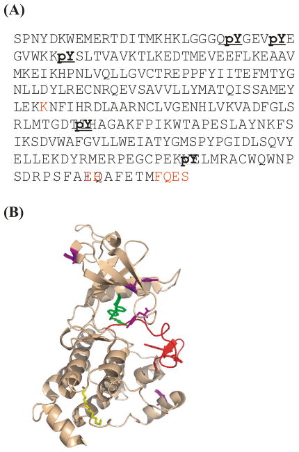
Figure 2.
Locating phosphorylation sites in Abl kinase via trypsin/subtilisin digestion and mass analysis. (A) Mass spectrometry identified phospho-tyrosine residues in primary sequence, marked in bold, in bacterial-expressed Abl kinase domain. Sequences undetected by mass analyses are marked in red. These seven are not subject to possible phosphorylation. (B) Locations of phosphorylated residues in its 3-D structure (PDB code 1OPJ). The active loop is colored red. All other phosphorylated sites are labelled in magenta.

