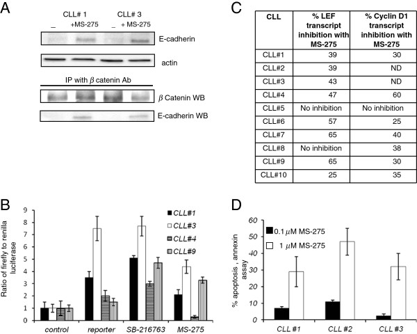
Figure 5.
Functional effects of HDACi on CLL specimens A. Two CLL specimens were treated with HDACi and western blot and immunoprecipitation analysis was performed. Top panel is an E-cadherin western blot with actin control showing the E- cadherin induction with HDACi exposure (MS-275 1.0μM, 48 hours). Bottom panel is the western blot analysis (WB) of the β-catenin immunoprecipitation material probed with anti-β-catenin antibody and anti-E-cadherin antibody. E-cadherin protein signal is observed in the beta-catenin immunoprecipitate material from HDACi treated CLL specimen. B. Wnt pathway luciferase reporter assay in CLL specimens. CLL specimens were transfected with the control or reporter constructs. Reporter transfected cells were separated in three groups, no treatment, SB-216763 compound or HDACi treatment. Data shown is a ratio of firefly to renilla. C. Table with real time PCR data with expression of LEF and cyclinD1 in CLL specimens treated with HDACi, MS-275. The numbers represent the percentage decrease in the expression of LEF and cyclinD1 in HDACi treated cells as compared to the non-treated control. D. Induction of apoptosis in CLL specimens with MS-275 treatment. Three CLL specimens were treated with two different concentration of HDACi MS-275 (0.1 and 1.0 μM concentration) and analyzed for induction of apoptosis at the 48 hour time point by a flow cytometry based annexin assay. (mean of three experiments).

