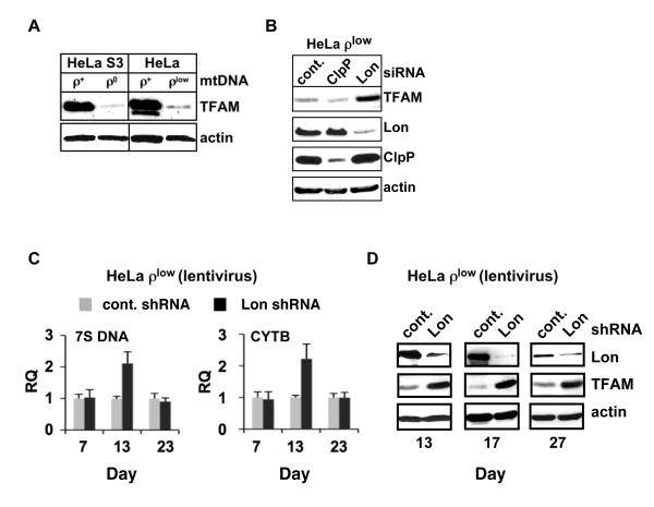
Figure 2. Lon knockdown in mtDNA-depleted cells increases TFAM and mtDNA.
(A) Extracts from HeLa ρ+, ρ0 and ρlow cells were blotted for TFAM. Overexposure permits detection of TFAM in ρ0 and ρlow cells. A lower molecular weight TFAM band in ρ+ cells is likely a processed form or breakdown product. (B) Extracts from ρlow cells transfected with control, Lon or ClpP siRNAs were blotted for TFAM, Lon, ClpP and actin. (C) Total DNA was isolated from ρlow cells transduced with control or Lon shRNA lentivirus (5 MOI) and relative quantitation (RQ) of mtDNA was determined by qPCR of 7S DNA and CYTB gene using the nuclear APP gene as an endogenous control. Data represent at least three independent experiments. Standard error of the mean is shown. (D) Extracts from ρlow cells transduced as in (C) were immunoblotted for Lon, TFAM and actin.

