
Figure 1.
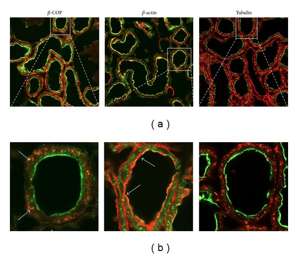
Confocal immunodetection in rat kidney cortex of NaPi-2a (green, Alexa488), β-COP (red, Alexa568), β-actin (red, phalloidin-rhodamine), and tubulin (red, Alexa 568). NaPi-2a colocalizes with β-COP intracellularly, and with β-actin in the BBM (see arrows, orange merge). NaPi-2a does not colocalize with tubulin (only green and red are visible, under steady-state conditions). (a) Low magnification of kidney cortex; (b) high magnification to show single proximal tubules.