Figure 6.

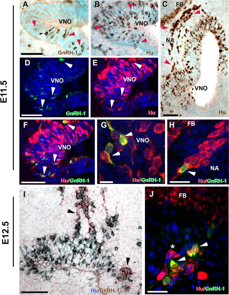
GnRH-1 cells emerge from the developing vomeronasal region as Hu-positive cells and increase GnRH-1 levels while migrating toward the forebrain junction (FBJ). Immunohistochemistry for GnRH-1 (A) and RNA binding protein Hu (B, C) on sections of the developing VNO at E11.5 show few GnRH-1-expressing neurons detectable in the VNO (A, red arrowheads). In contrast, large numbers of neuronal precursors (Hu positive) were present both within the VNO (B, red arrowhead) and in the nasal region (NA) extending from the VNO toward the forebrain (FB) (C). D–H, Double immunofluorescence for GnRH-1 (green) and Hu (red). D–F, In the developing VNO at E11.5, a few Hu-positive neuronal precursors start to express GnRH-1 (arrowheads in D, E; merge in F). G, High magnification (100×) within the VNO anlage showing Hu-positive/GnRH-1-positive GnRH-1 neuron precursors (arrowheads). H, Hu-positive cells (red) migrating toward the forebrain (see C for localization), Hu/GnRH-1 double-positive maturing GnRH-1 neuron (arrowhead). I, Double immunocytochemistry for Hu (blue) and GnRH-1 (brown) at E12.5 showed large groups of Hu-positive and maturating Hu-positive/GnRH-1-positive neurons (arrowheads) migrating out of the VNO. J, Double immunofluorescence for Hu (red) and GnRH-1 (green) at E12.5 highlighted maturing Hu/GnRH-1 neurons (arrowhead) in proximity of the forebrain as well as Hu single-labeled migratory neurons (*). Scale bars: A–F, I, 50 μm; H, J, 20 μm; G, 10 μm.
