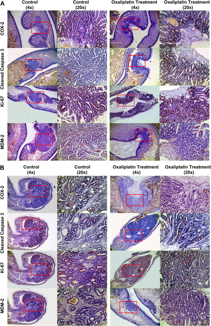
Fig. 6.
Immunohistochemical analysis of adenomas found in the small intestine (A) and colon (B) stained for Cox-2, Cleaved Caspase-3, Ki-67 and Mdm-2. Each panel compares adenomas found in the control and treatment group. Section magnified is selected in a red rectangle and stained cells are pointed out with red arrows. All shown sections are at 4× magnification (bar = 1000um) and further magnified to show details at 20× (bar = 100 um). Immunohistochemistry was performed using Cox-2 (Santa Cruz, CA), Cleaved Caspase-3 (Cell Signaling), Ki-67 (Dako) and Mdm-2 (Santa Cruz, CA) antibodies at a 1:500 dilution. Immunohistochemical signal was detected by secondary biotinylated goat anti-mouse antibody (1:500 dilution; Santa Cruz, CA) followed by Vector ABC tertiary kit (Vector Laboratories, Burlingame, CA, USA) according to the manufacturer’s instructions. All immunohistochemistry was performed on a BondMax machine (Vision Biosystems, Newcastle, UK) according to the manufacturer’s instructions and involved antigen retrieval by BondMax Epitope retrieval solution heated on the machine for 20 min. (For interpretation of the references to colour in this figure legend, the reader is referred to the web version of this article.)

