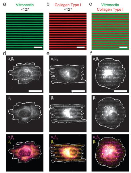
Fig. 3. Integrin segregation on multicolor surface patterns.
(a)–(c) Fluorescent micrographs of BSA conjugated to AlexaFluor-488 (green) to represent vitronectin, BSA conjugated to AlexaFluor-594 (red) to represent collagen type I and black to represent non-adhesive F127. (d)–(f) Micrographs of human umbilical vein endothelial cells (HUVECs), seeded on substrates patterned as in (a)–(c), fixed after 1 h, and immunolabeled. Note the colocalization of αvβ5 integrin to vitronectin, and β1 integrin to collagen type I. All scale bars, 20 μm.

