
Fig. 4. Continuous adhesions composed of compositionally and spatially segregated types of integrins.
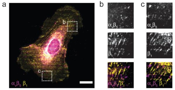
(a–c) Micrograph of a HUVEC seeded on a pattern with alternating lines of vitronectin and collagen type I as in Fig. 3c, fixed 2 h after seeding and immunolabaled. Panels (b) and (c) correspond boxed regions in (a). Note the segregation of αvβ5 and β1 integrins throughout the cell (a), and within single adhesions (b, c). Scale bar, 20 μm.