
Figure 1.
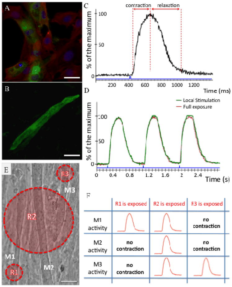
Generation and light-induced stimulation of ChR2-expressing skeletal muscle cells in vitro. (A) Fluorescence image of the membrane-bound ChR2-GFP signal (green) overlaid with F-actin immunostaining (red) in C2C12 myoblasts. Nuclei are shown in blue. (B) Multi-nucleated myotubes expressing the GFP signal (green). (C) Typical contraction pattern of a ChR2-GFP expressing myotube upon stimulation with blue light pulses (10 mW mm−2) for durations indicated by blue bars. A representative example of 20 experiments is shown. (D) Representative repetitive contraction pattern evoked by local and full exposure of a ChR2-GFP–expressing myotube with blue light pulses (10 mW mm−2) for durations indicated by blue bars. (E) Selective activation of myotubes with local stimulation. Myotubes are denoted by M1, M2 and M3 and regions of exposure are labeled with R1, R2, and R3. (F) Blue light pulses confined to a region stimulate only the myotubes residing inside that specific region. (Scale bars: A, 10 μm; B and E, 20 μm).