
Figure 4. NPSLE with Thrombotic and Cerebral Calcinosis.
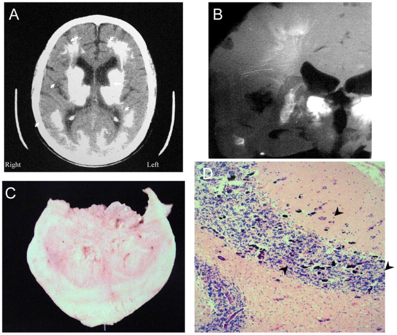
Figure 4 shows further features of the same individual as in Figure 3 (Subject 2). Figure 4A. The computed tomographic (CT) image demonstrates extensive calcification in the thalamus, putamen, caudate nucleus, white matter, and posterior gray matter (arrowheads). Figure 4B. A radiograph of the brain slice at autopsy shows lacey linear and flowering calcifications that follow the arteriolar and venular vasculature, as well as complete calcification of the caudate nucleus (arrowhead). Figure 4C shows one of the calcific spheroids after excision. Figure 4D demonstrates extensive microcalcifications at the gray matter-white matter junctions in the cerebellum (arrowheads; H&E stain, Magnification X 100).