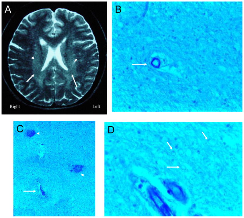
Figure 5. NPSLE with Accelerated SLE Activity and Confusional State.

Figure 5A (Subject 3) is a T2-weighted MRI demonstrating vague periventricular white matter hyperintensities (arrowheads), foci of reduced signal in white matter, and scattered focal white matter lesions. Histological changes consisted of ill-defined areas of pallor commonly accentuated in the immediate vicinity of blood vessels and showing average reduction in numbers of oligodendrocytes with generally non-thrombosed blood vessels (Figure 5B, arrow; LFB/PAS stain, Magnification X 150). Figure 5C. In each putamen and left thalamus multiple small areas of necrosis (arrow), frequently hemorrhagic, contained ischemic neurons and showed mild gliosis and sparse macrophages at their periphery with frequent microhemorrhages (arrowheads; LFB/PAS stain, Magnification X 100). Figure 5D. The density of myelinated fibers was diminished and under high magnification, the fibers had a beaded appearance (arrows; LFB/PAS stain, Magnification X 200). The intrinsic and extrinsic cerebral blood vessels showed no abnormalities.
