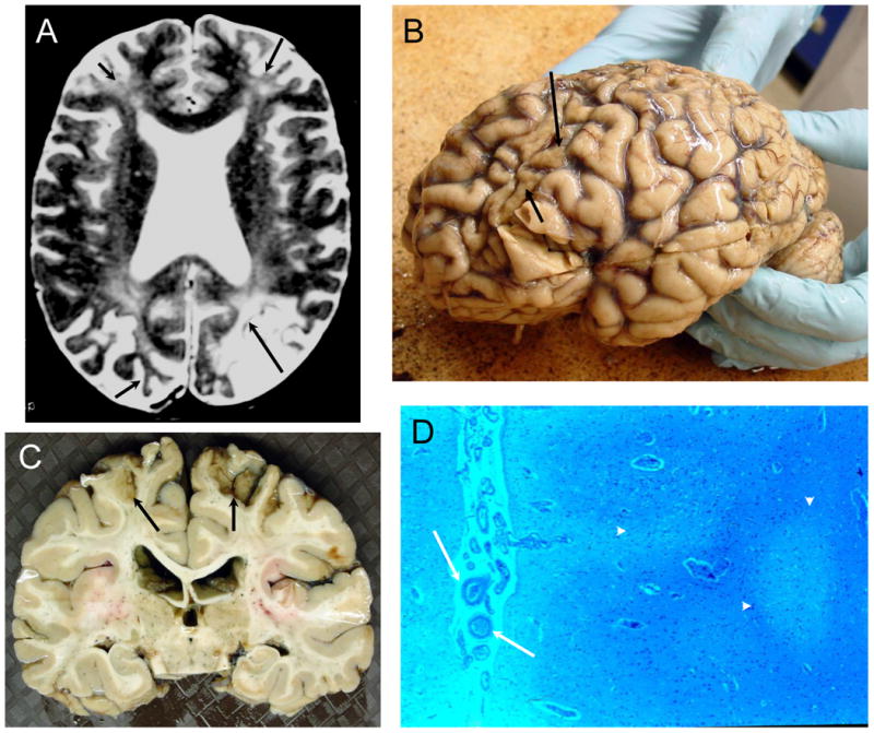
Figure 6. NPSLE with Chronic Thrombotic Cerebrovascular Disease.

Figure 6A (Subject 4). The T2-weighted MRI demonstrates severe cortical atrophy, severe ventricular dilation, multiple small focal white matter abnormalities, diffuse hyperintensities in deep white matter, and old cortical infarcts with focal atrophy in both frontal lobes, left occipital lobe, and right occipital lobe (arrows). Figure 6B (Subject 4). The frontal lobes were flattened, and the cerebral gyri demonstrated sickle-shaped atrophic post-ischemic bands in the frontal and occipital areas (arrows) with evident generalized atrophy. Figure 6C (Subject 4). The brain slice demonstrates the focal frontoparietal post-ischemic atrophy superiorly (arrows). Figure 6D (Subject 4). The cortex is unevenly depleted of neurons with evident atrophy (arrowheads), sometimes laminar in distribution, with increases in astrocytes nuclei and adjacent reductions in white matter and axons with some glial hyperplasia and evidence of thromboembolic vasculopathy (arrows; LFB/PAS stain, Magnification X 50).
