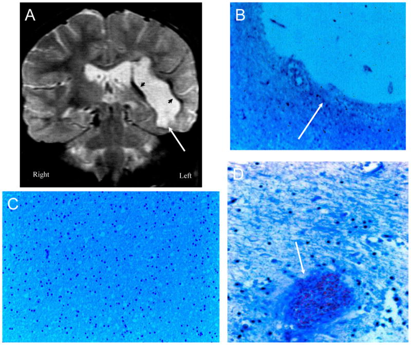
Figure 9. NPSLE with Accelerated SLE, Hypertension, and Intra-cerebral Hemorrhage.

Figure 9A (Subject 8). The T2-weighted MRI demonstrates a large resolved intracerebral hemorrhage of the right hemisphere with formation of a cerebrospinal fluid filled cyst that appears as a hyperintense fluid mass next to the ventricles (arrow). A thin dark rim of hypointensities around the cyst represents dense connective tissue and hemosiderin-laden macrophages (arrowheads). Figure 9B (Subject 8). The cyst is lined with cortical tissue with marked gliosis and hemosiderin-laden macrophages (arrow; LFB/PAS stain, Magnification X 100. Figure 9C (Subject 8). The remainder of the cortex is relative unremarkable (LFB/PAS stain, Magnification X 100). Figure 9D (Subject 12). Sections of the corpus striatum demonstrate focal perivascular siderophages without vasculitis, and satellite hemorrhages (arrow) and edema in the parenchyma surround the hematoma (LFB/PAS stain, Magnification X 100).
