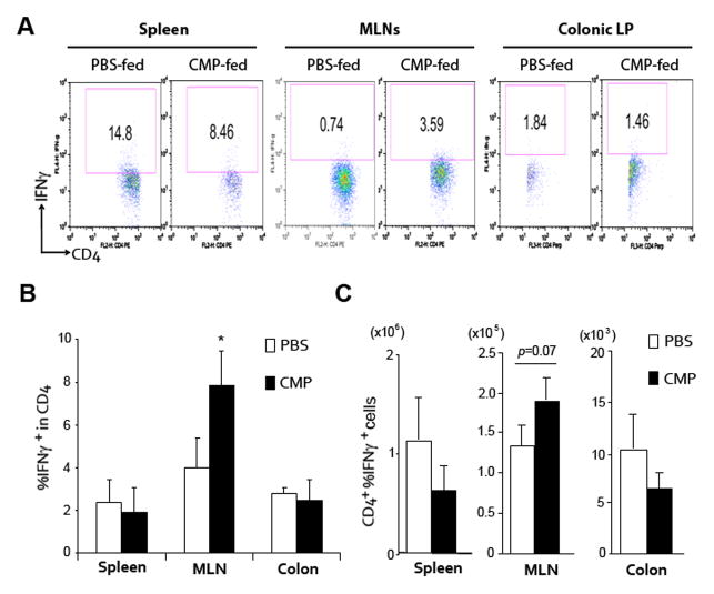
Figure 4.
CMP-treatment significantly enhances IFNγ production in a DSS-induced colitis model. (A–C) Cells in spleen, mesenteric lymph nodes (MLNs) or colonic lamina propria (LP) of 4% DSS-treated WT mice on Day 12 were stained with FITC-anti CD4 and APC-IFNγ, antibodies, and were assessed by flowcytometry as shown in A. The Numbers indicate the percent of CD4 and IFNγ double-positive cells within the indicated gates (A). The numbers of percent IFNγ positive cells within the total CD4+ T cells (B) or absolute number of CD4 and IFNγ double-positive cells (C) in each group of mice are shown. *P <.05 (CMP-treated versus PBS-treated mice in each tissue).

