
Fig 1.
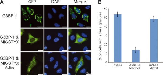
MK-STYX inhibits G3BP-1 induced stress granules. (A) Representative examples of the subcellular distribution patterns of G3BP-1. HeLa cells cotransfected with expression vectors for G3BP-1 and wild-type MK-STYX showed fewer cells with stress granules. Overexpression of G3BP-1 and MK-STYX active mutant resulted in intermediate stress granule assembly. Merged images show the location of GFP-tagged G3BP-1 (green) relative to DAPI-stained nuclei (blue). Scale bar = 10 μm. (B) HeLa cells were cotransfected with expression vectors for G3BP1-GFP and either MK-STYX or MK-STYX active mutant. Cells were analyzed 24 h post-transfection for G3BP-induced stress granule assembly by fluorescence microscopy, after staining with DAPI to reveal the nuclei. Cells were scored for the presence or absence of stress granules. Three replicate experiments were performed (n = 100); error bars indicate the SEM.