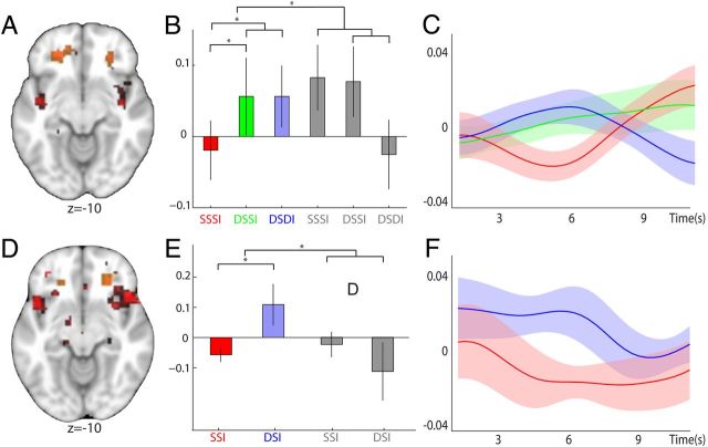
Figure 3.
Representations of stimulus–reward associations. A, D, A bilateral region in lOFC was found to represent stimulus–reward information for food rewards. Trials with repeated elicitation of the same stimulus–reward mapping (SSSI) were compared against trials with two different predictive stimuli (DSSI and DSDI). To control for effects of mere visual stimulus adaptation, trials with food items were further contrasted against trials with neutral items (A) (((DSDIf + DSSIf) − (2 × SSSIf)) − ((DSDIn + DSSIn) − (2 × SSSIn))). In a similar way, we also analyzed between-trial adaptation effects by contrasting all trials that were preceded by a common stimulus–reward mapping, to those in which the stimulus–reward pairing of the preceding trial differed (D) ((DSIf − SSIf) − (DSIn − SSIn)). Regions demonstrating adaptation to stimulus–reward encoding in both the within- and between-trial contrast are shown (both p < 0.05 for visualization). This reveals that for both contrasts a region in rostro-lateral OFC adapted to the pairing of stimulus–reward information (shown in red); the extracted ROIs are overlaid for visualization and are shown in orange. B, C, E, F, Parameter estimates and time courses are shown as in Figure 2 (red, SSSIf; green, DSSIf; blue, DSDIf; gray, SSSIa, DSSIa, and DSDIa). B, C, Adaptation in lOFC to stimulus–reward information within trials, extracted from an orthogonal ROI defined by the equivalent between-trial contrast. E, F, Similar adaptation effects between trials in lOFC, extracted from the orthogonal within-trial ROI.

