Fig. 2.
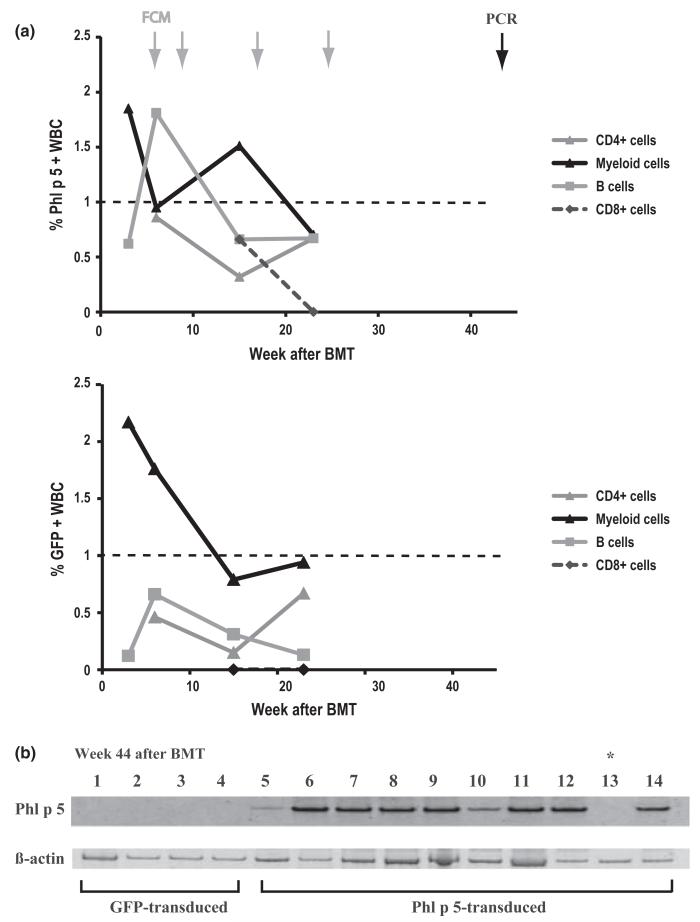
Phl p 5-chimerism is detectable in flow cytometry at early and PCR at late time points. (a) Phl p 5-specific expression within lineages of WBC as indicated was detected by flow cytometry at early time points. Data represent the mean% of Phl p 5 chimerism in recipients of Phl p 5-transduced BM (n = 10). Arrows in grey indicate time points of FCM analysis, arrow in black indicates PCR analysis at 44 weeks post-BMT. Note: Phl p 5-expressing CD4+ and CD8+ cells became detectable only 6–15 weeks after BMT due to delayed recovery of T cells after administration of T-cell-depleting antibodies (b) Upper gel: Phl p 5-specific PCR-products (lane 1–4) shown in genomic DNA of recipients transplanted with GFP-transduced BM (GFP-transduced) and recipients of Phl p 5-transduced BM (Phl p 5-transduced; lane 5–14). In chromosomal DNA of mouse 9 (lane 13) no Phl p 5-specific product was detectable (*). Lower gel: Lane 1–14 show β-actin specific PCR-products in genomic DNA of splenocytes of recipients of GFP-transduced BM (lane 1–4) and Phl p 5-transduced BM (lane 5–14).

