
Figure 4.
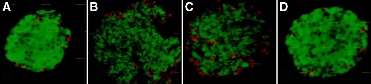
Pancreatic islet morphology at different stages of infection. Immunohistochemical analysis of pancreatic islets. Insulin-stained β-cells are green, and glucagon-stained α-cells are red. A: Classical morphology at baseline. B: There was a dramatic disruption of islet integrity at day 15 postinfection. C: The disruption of islet integrity continued at day 30 postinfection. Note the disorganized structure of the α-cells on day 30, which are usually located in the periphery of mouse islets. D: There appears to be partial recovery of islet integrity at day 100 postinfection.