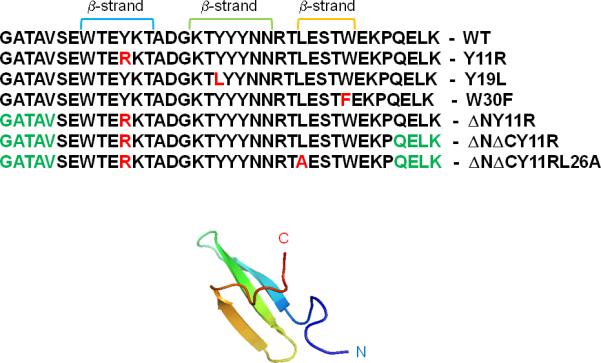
Figure 1.

Experimental NMR structure of triple β-strand WW domain from the Formin binding protein 28 (FBP) [PDB: 1E0L]. The sequences of the WT, all full-size and truncated mutants of FBP28 WW domain with mutated residues (in red color) and ΔN/ΔC truncations (in green color), are shown.
