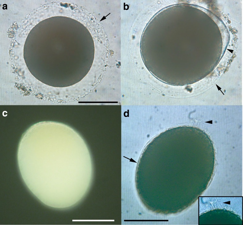
Figure 1. Eggs and hatchling of Xenoturbella bocki.
(a) Unfertilized egg with a transparent layer (arrow). (b) Embryo before hatching within the fertilization envelope (arrowhead), surrounded by the transparent outer layer (arrow). (c) Embryo a day after hatching with anterior end at top left. (d) Embryo 3 days after hatching, uniformly covered with cilia (arrow). Apical tuft (arrowhead) at top right, also shown enlarged in inset. Scale bars, 100 μm.

