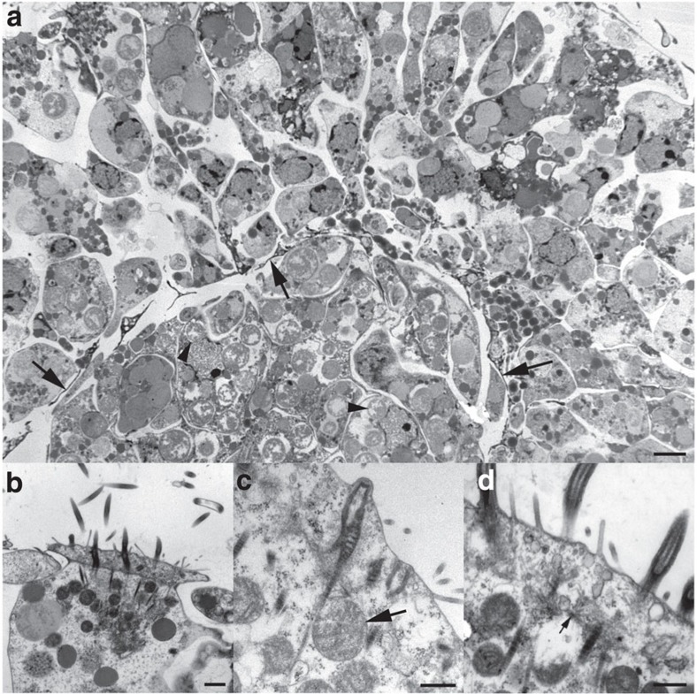
Figure 3. Transmission electron microscopy of 4-day free-swimming hatchling of Xenoturbella bocki.
(a) Cross-section of the body. Note the thin electron-dense cytoplasmic extensions of muscle cells, delimiting the outer epidermal layer from the inner gastrodermal cell mass (arrows). Many rounded bodies, presumed to be symbiotic bacteria, are present in the gastrodermal cells (arrowheads). Scale bar, 2 μm. (b) Surface of a multiciliated epidermal cell with ciliary axonemes and double vertical rootlets. The epidermal cells are laterally interdigitating. Scale bar, 1 μm. (c) Epidermal cell surface with double ciliary rootlets in longitudinal section. Note mitochondria (arrow) close to rootlets. Scale bar, 0.5 μm. (d) Epidermal cell surface with ciliary rootlets. Note microtubuli spreading out from the ciliary basal foot (arrow). Scale bar, 0.5 μm.

