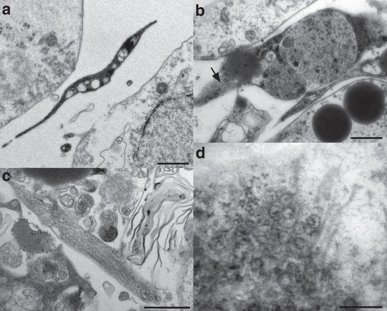
Figure 4. Transmission electron microscopy of 4-day free-swimming hatchling of Xenoturbella bocki.
(a) Cytoplasmic extension of developing muscle cell, situated between the epidermal cell layer and the gastric cell mass. Scale bar, 0.5 μm. (b) Developing muscle cell with electron-dense cytoplasm and myofilament bundles in cross-section, also seen in oblique section (arrow). Scale bar, 0.5 μm. (c) Part of developing muscle cell, showing myofilaments in longitudinal section. Scale bar, 0.5 μm. (d) Point-core neuronal vesicles and neurotubules in nerve cell synapse. Scale bar, 0.25 μm.

