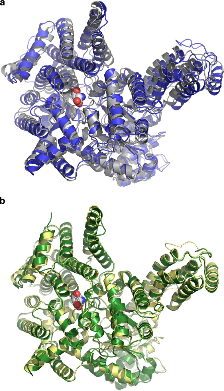
Figure 1. Structural comparison of C3 PEPC and C4 PEPC monomers.
(a) Overlay of PEPC from E. coli (C3-type, grey)17 and Z. mays (C4-type, blue)17. The calculated root-mean-square deviaton (r.m.s.d.) of both monomers is 2.0 Å. (b) Superposition of PEPC from F. pringlei (C3-type, green) and F. trinervia (C4-type, yellow). The Flaveria PEPCs have a high overall topological similarity with a r.m.s.d. of 0.4 Å. Structures are shown as ribbons with the aspartate/malate-binding site on top. The bound inhibitors are shown as spheres representing van der Waals radii.

