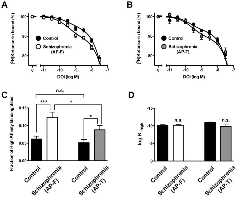
Fig. 5.
(A) [3H]Ketanserin specific binding (2 nM) displacement curves by DOI in postmortem frontal cortex of antipsychotic-free (AP-F) schizophrenic subjects and controls. (B) [3H]Ketanserin specific binding (2 nM) displacement curves by DOI in postmortem frontal cortex of antipsychotic-treated (AP-T) schizophrenic subjects and controls. (C) Fraction of high-affinity sites of DOI displacing [3H]ketanserin specific binding. (D) Normalized high-affinity (log Ki-high) of DOI displacing [3H]ketanserin specific binding in schizophrenic subjects and individually matched controls. *p<0.05; ***p<0.001; n.s., not significant; Student’s t-test. See Supplementary Tables 1 and 2 for demographic information. See Table 1 for statistical analysis. All values represent means±SEM.

