Figure 2.

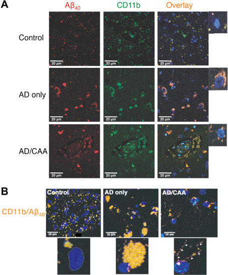
Representative confocal micrographs of fixed human occipital lobe tissue. Images were taken at 630× magnification and immunostained for (A) CD11b (n = 4 for each group) and colocalized with an anti‐Aβ40 antibody. A FITC‐conjugated secondary antibody was used for CD11b (green) and a Texas Red‐conjugated secondary antibody was used for Aβ40 (red). Nuclei were stained with DAPI (blue). (B). Representative Zstack images of microglia colocalized for CD11b and Aβ40. Panels show colocalized images in which microglial nuclei are stained with DAPI and colocalization of CD11b and Aβ40 is stained yellow/orange. Note the colocalization of CD11b with Aβ40 in punctate densities at the plasma membrane in AD/CAA (arrowheads), rather than the perinuclear, phagocytic vesicle distribution as seen in AD‐only cases. Controls show only faint colocalization for CD11b and Aβ40 that appear to localize to microglial processes.
