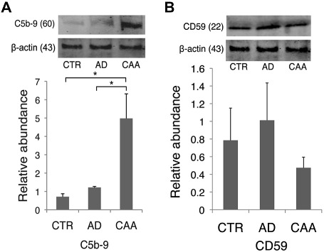
Figure 4.

Western blot for C5b‐9 and CD59 on isolated cerebral blood vessels. A. C5b‐9 was significantly increased on isolated AD/CAA vessels compared to both controls and AD‐only vessels. B. CD59 showed a trending decrease on occipital blood vessels of AD/CAA cases compared to the control and AD‐only groups. *P < 0.05; **P < 0.01. Data represented as mean ± SEM. Relative abundance is the ratio of C5b‐9 and CD59:beta‐actin as optical density.
