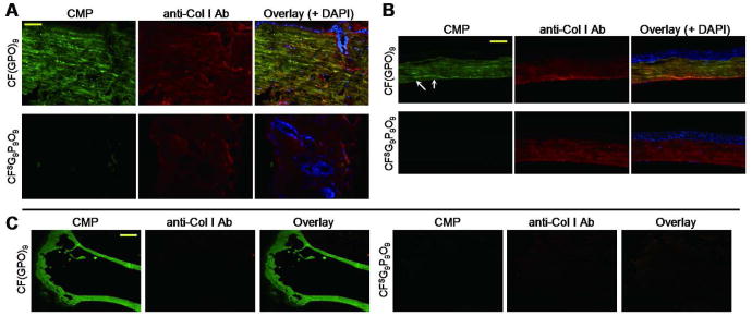
Figure 4.
Histological tissue staining using photo-activated caged CMPs. Fluorescent micrographs of fixed mouse skin (A) and cornea (B) sections probed by photo-triggered CFNB(GPO)9 (in green, top panels) or scrambled peptide CFSG9P9O9 (in green, bottom panels) and co-stained with anti-collagen I antibody (red), and DAPI (blue). The CFNB(GPO)9 staining revealed fine irregular collagen fibers of the dermis (A) and parallel collagen fibrils in the corneal stroma as well as the collagens in Descemet's membrane (arrows) (B). (C) Fluorescent images of paraffin embedded, demineralized mouse tibia sections stained with photo-triggered CFNB(GPO)9 (green, left panels) or scrambled peptide CFSG9P9O9 (green, right panels), and co-stained with anti-collagen I antibody (red), showing prominent CMP signals and only weak non-specific antibody signals from the collagenous bone. Concentrations of CFNB(GPO)9 and CFSG9P9O9 used in this study are: (A), 25 μM; (B), 2.5 μM; (C), 8 μM. (scale bars: A, 50 μm; B, 100 μm; C, 0.5 mm).

