
Fig. 2. Histopathological findings in C9ORF72 expansion carriers.
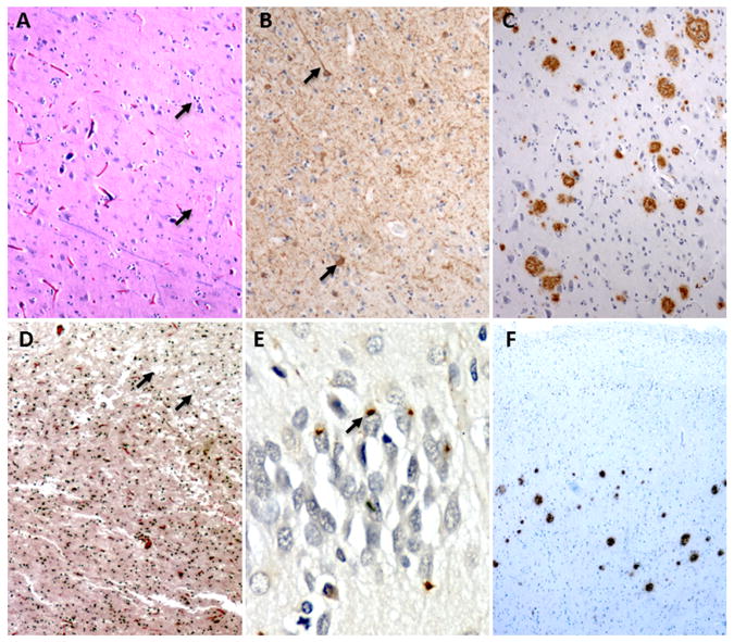
(A – C) The index case in family 6 showed classic AD changes including (A) neuronal loss and plaques (B) tau positive threads and tangles, and (C) amyloid plaques throughout the neocortex and hippocampus. (D – F) The index patient from family 2 showed (D) microvacuolation in the frontal cortex, (E) ubiquitin-immunoreactive intracellular inclusions in the hippocampus, and (F) cortical extracellular amyloid plaques. For more details see supplementary data.