Figure 4.
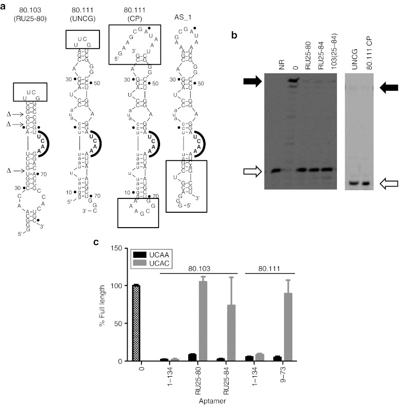
Utilization of UCAA bulge and peripheral flanking sequences. (a) Sequences and projected secondary structures of some of the aptamer variants analyzed here. Boxed portions indicate segments addressed by individual mutants. (b) Two gels showing results of simplifying aptamers 80.103 (left) and 80.111 (right). The “RU” designation indicates that three internal unpaired nucleotides were removed from the corresponding truncations of aptamer 80.103. 80.111 mutants “UNCG” and “CP” are shown in panel A. Open and filled arrows indicate positions of unextended primer and full-length product bands, respectively. Results from additional internal mutations are given in Supplementary Figure S6. (c) Partial disruption of UCAA element. Primer extension by RT was measured in the presence of several variants of aptamers 80.103 and 80.111. Black bars indicate parental sequences. Gray bars indicate mutated sequences in which UCAA bulges were mutated to UCAC. Error bars indicate the SDs among three independent measurements. NR, no reaction.

