
Figure 4.
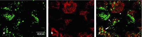
Double immunofluorescence with antibodies against tubulinIII. To specify the transduced cell types, (a) double immunolabeling with antibodies against β-galactosidase (coupled to Alexa-488, green) and (b) against the neuronal marker protein tubulinIII (coupled to Alexa-594, red) was performed. (c) Merge picture shows that almost all β-galactosidase expression occurs in cells staining positive for tubulinIII (see arrows), indicating that most of the transduced cells are neurons.