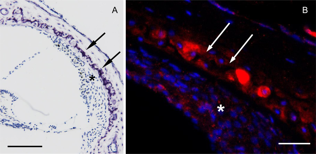
Figure 3.
Bmpr1b localizes to cartilaginous cell rests of the otic capsule. Cochlear cross section stained with azure (A), and with anti sense probe for bmpr1b (B). The red fluorescent signal (B) is most intense in cartilaginous rests (double arrows) and weaker in the spiral ligament (asterisk). Cell nuclei are in blue. Scale bar = 100 µm (A), 50 µm (B).

