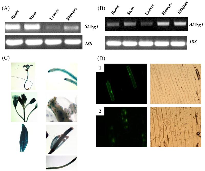
Fig. 2.
Tissue-specific expression and sub-cellular localization of Asg1. (A) and (B) expression of Asg1 in different tissues of potato and Arabidopsis, respectively; (C) GUS staining of transgenic Arabidopsis plants expressing the reporter gene β-glucoronidase driven by the promoter of AtAsg1. Leaves, roots, siliques, flowers and stems were stained. 5-Day old seedlings were also stained; (D) subcellular localization of the protein encoded by StAsg1. Confocal microscopy visualization of onion cells expressing GFP (1) and the fusion protein ASG1-GFP (2) 48 h after the transformation. Left panels, visualization at 490 nm. Right panels, bright field image.

